Professional USB microscope · Podiatry
Podiatry
Examination of nails, plantar skin, warts, and foot lesions for assessment and follow-up.
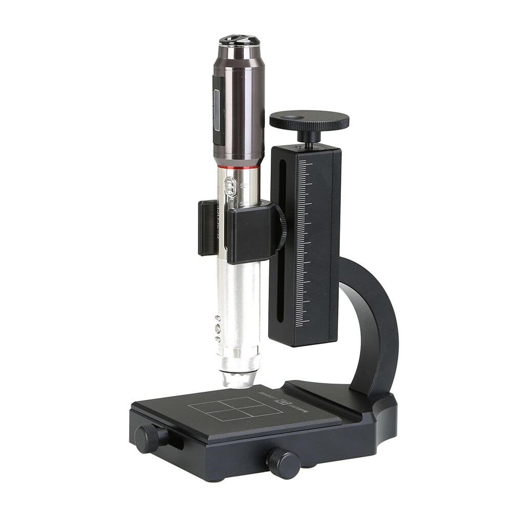
- Four quick magnification levels40x, 100x, 180x and 250x with the general inspection end-cap
- 3Pi narrow-tip end-cap100x access to small or recessed areas that remain in direct line of sight
- Quality optics, autofocus + Full HDCorning® Varioptic® liquid lens, photos and 30fps video
- Professional European supportShips from Spain with a two-year warranty
Overview
Podiatrists face a wide range of nail and skin conditions that require detailed visualization. Smart G-Scope helps examine onychomycosis, ingrown nails, hyperkeratosis, plantar warts, and vascular lesions with the detail needed for a clinically oriented assessment.
Longitudinal documentation can support clinician-led follow-up, including diabetic or at-risk feet, without replacing examination, vascular assessment, or diagnostic tests.
Images can be shared with dermatologists or vascular surgeons when interdisciplinary assessment is needed, improving care coordination.
Key benefits
- Detailed visualization of nail pathology
- Documentation of hyperkeratosis and plantar lesions
- Follow-up of antifungal treatments
- Consistent image documentation for clinician-led at-risk foot follow-up
- Improved visual communication with other specialists
Image gallery
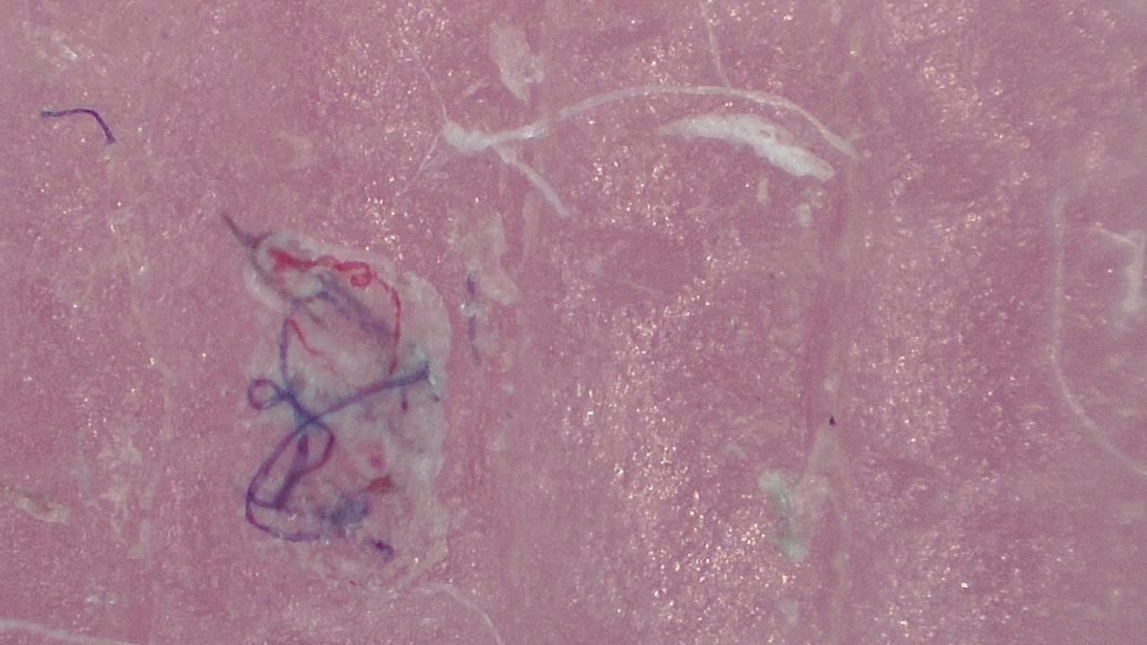
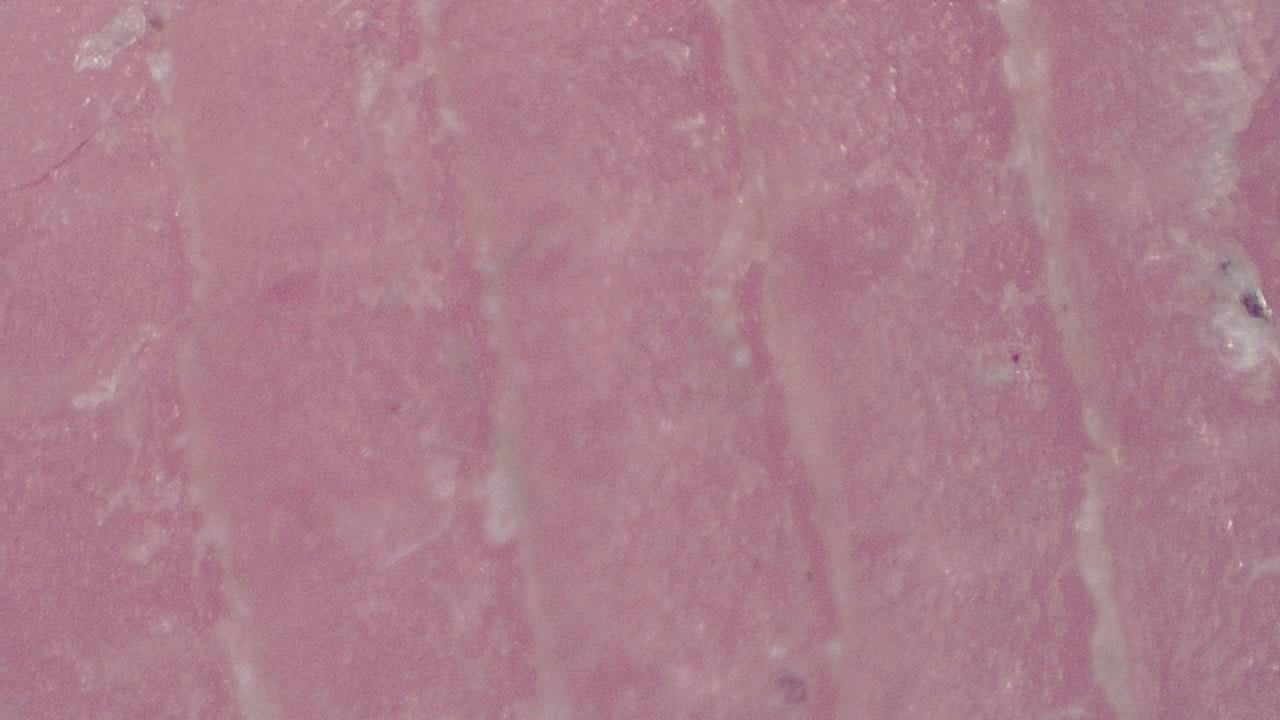
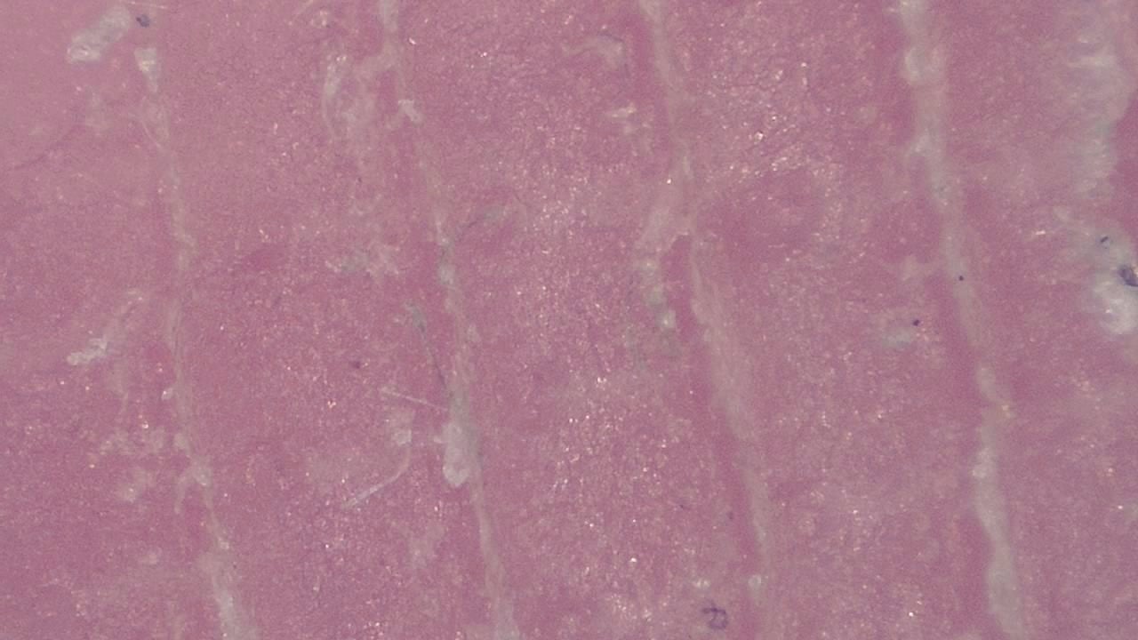
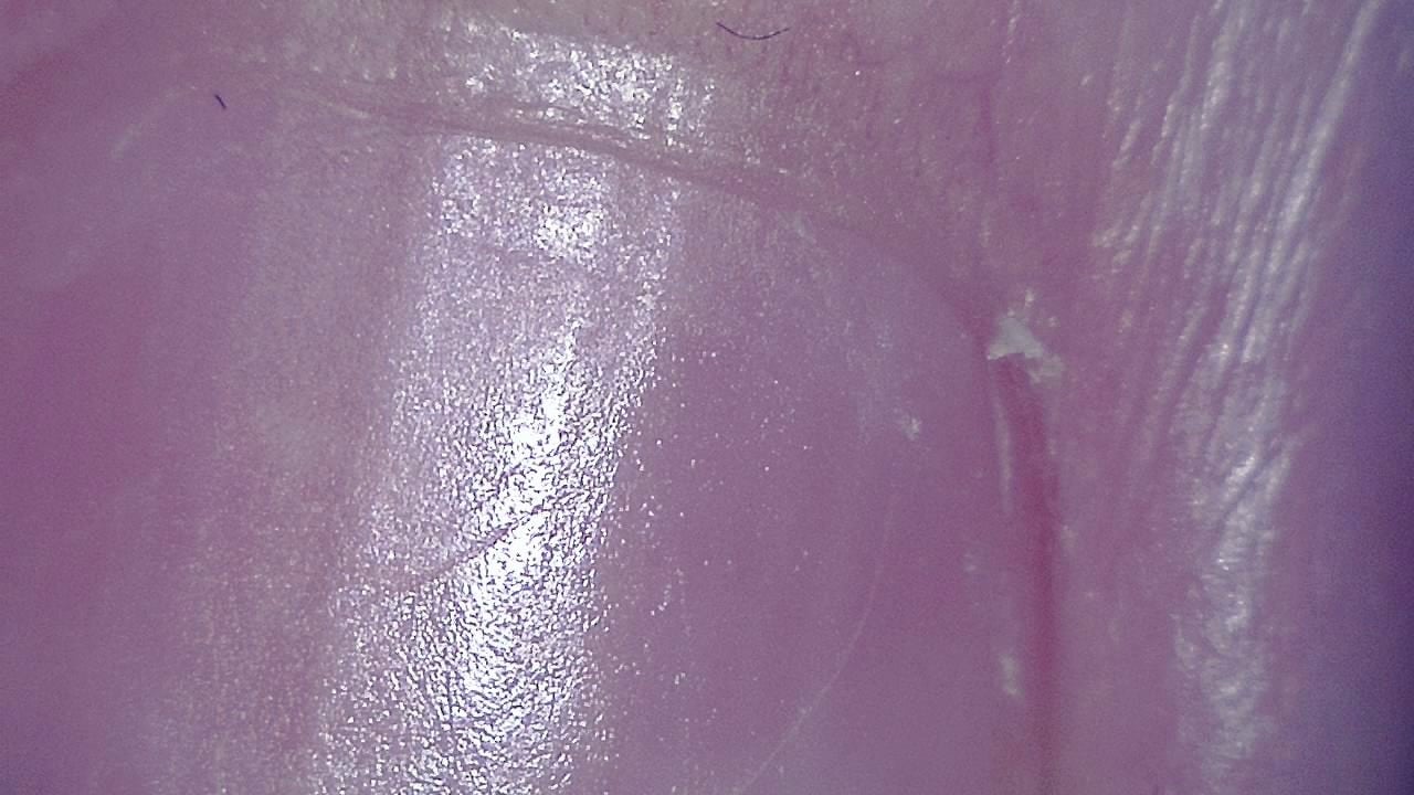
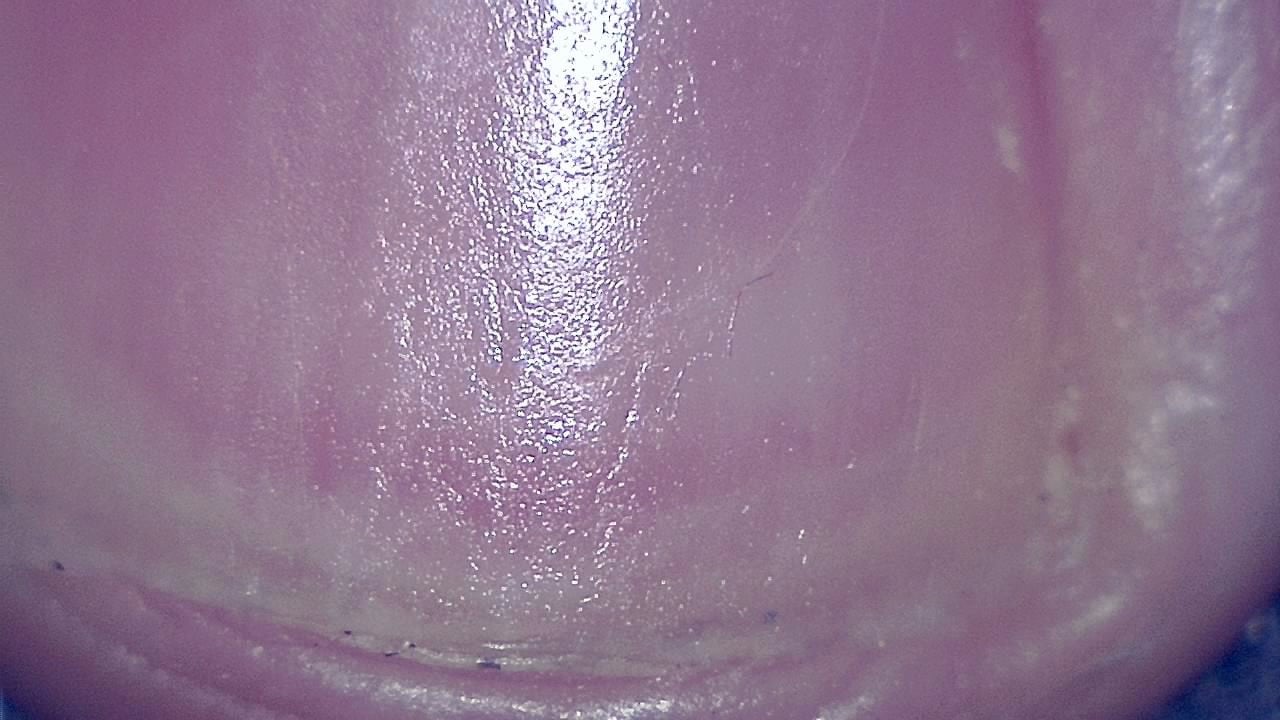
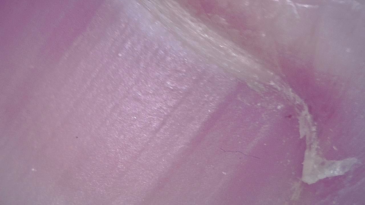
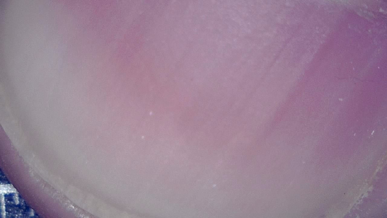
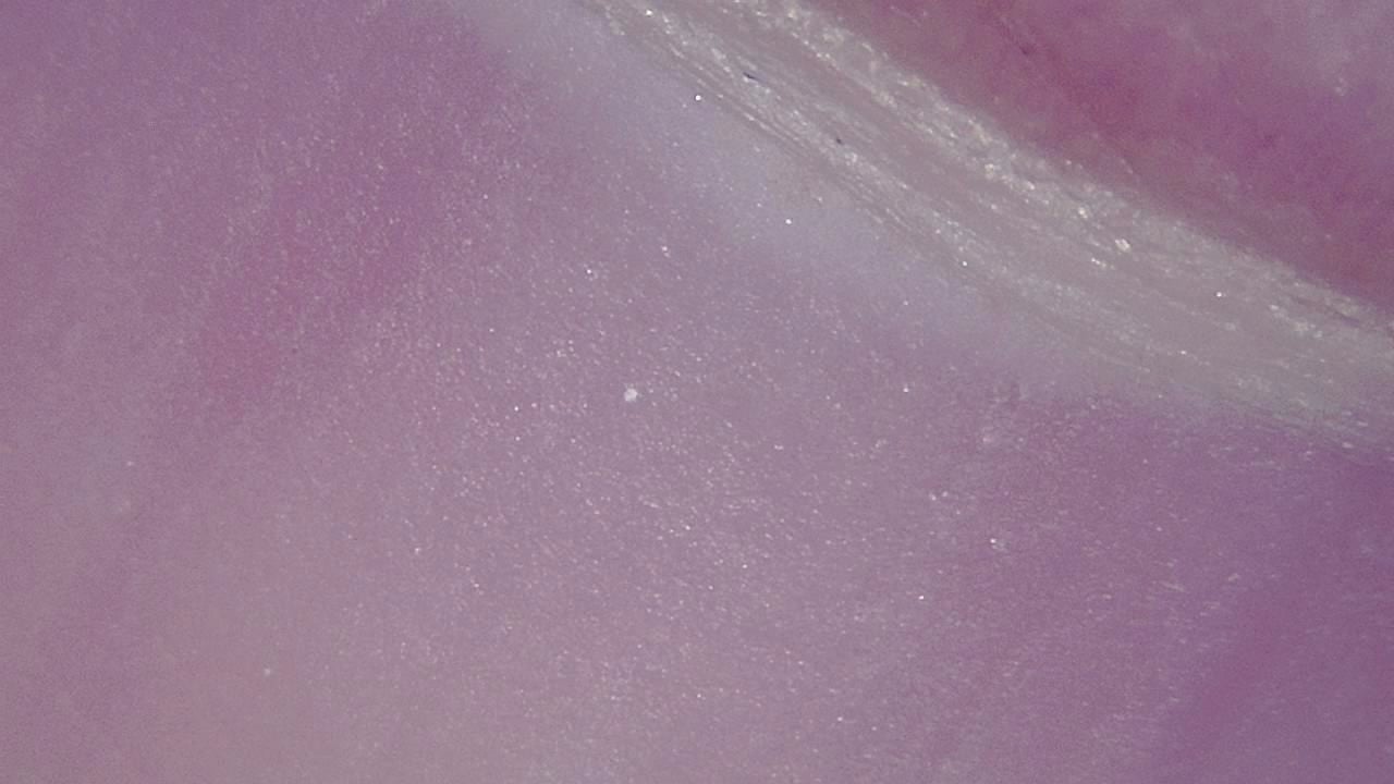
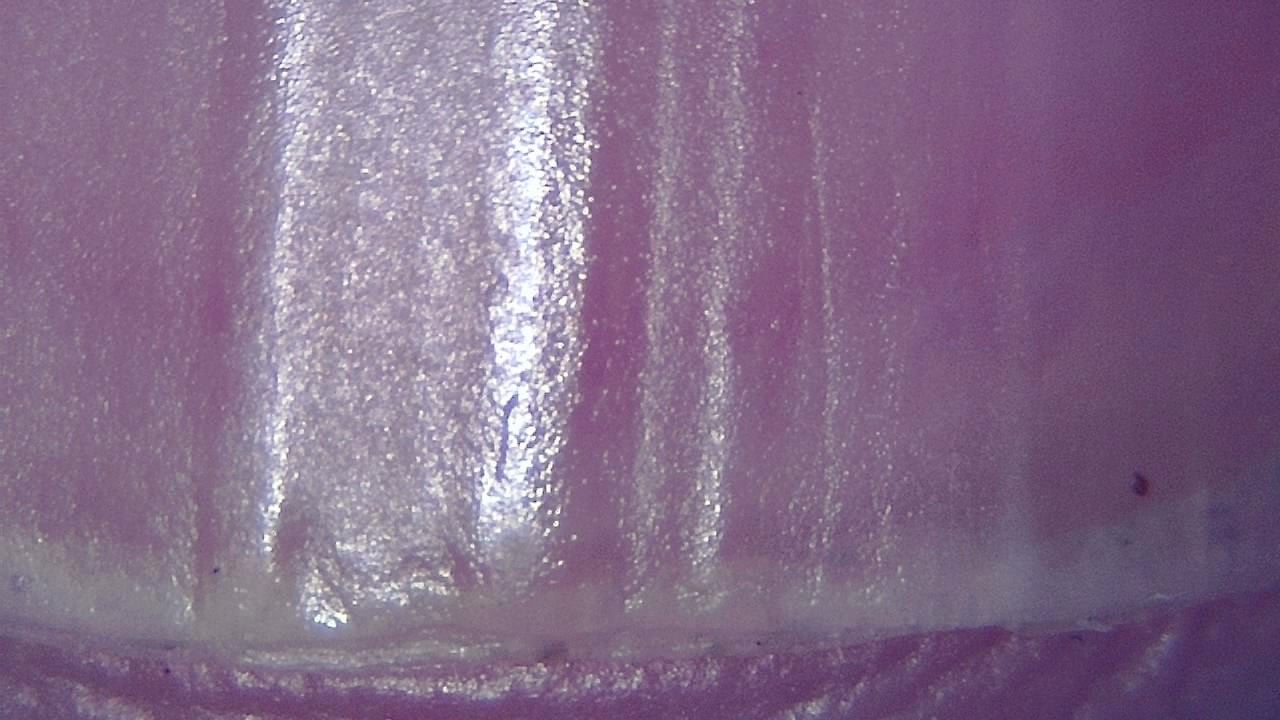
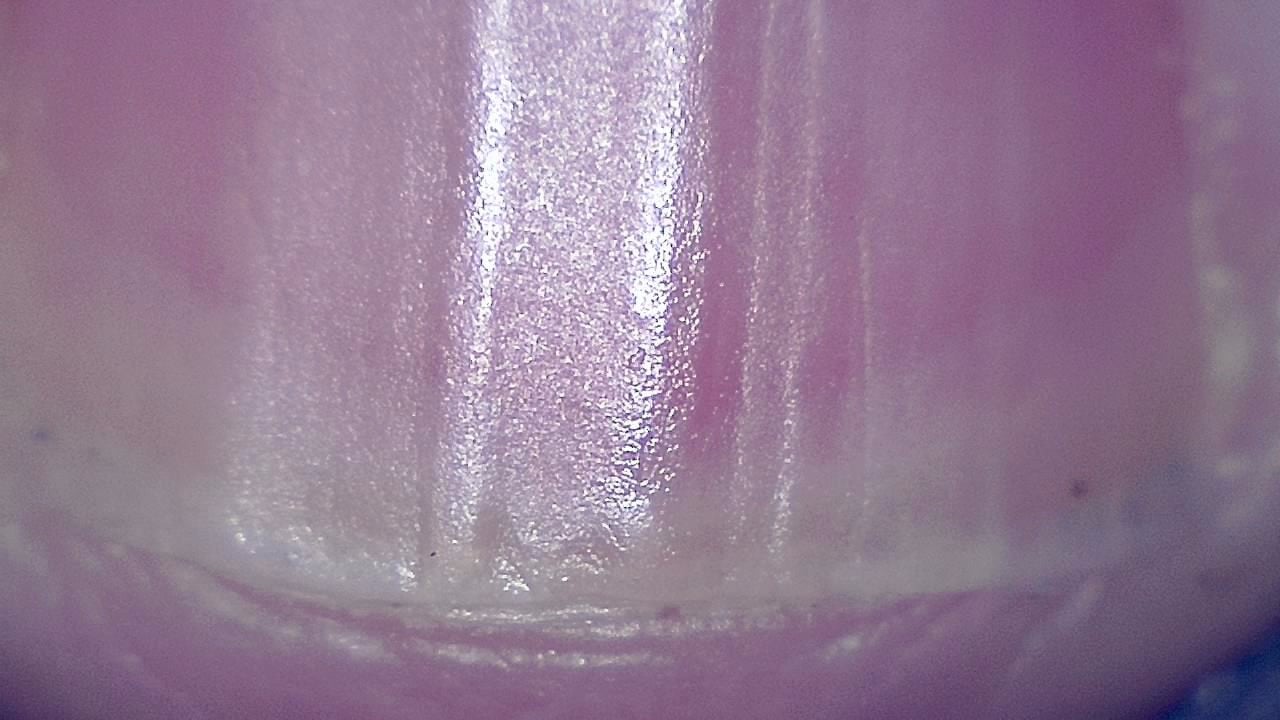
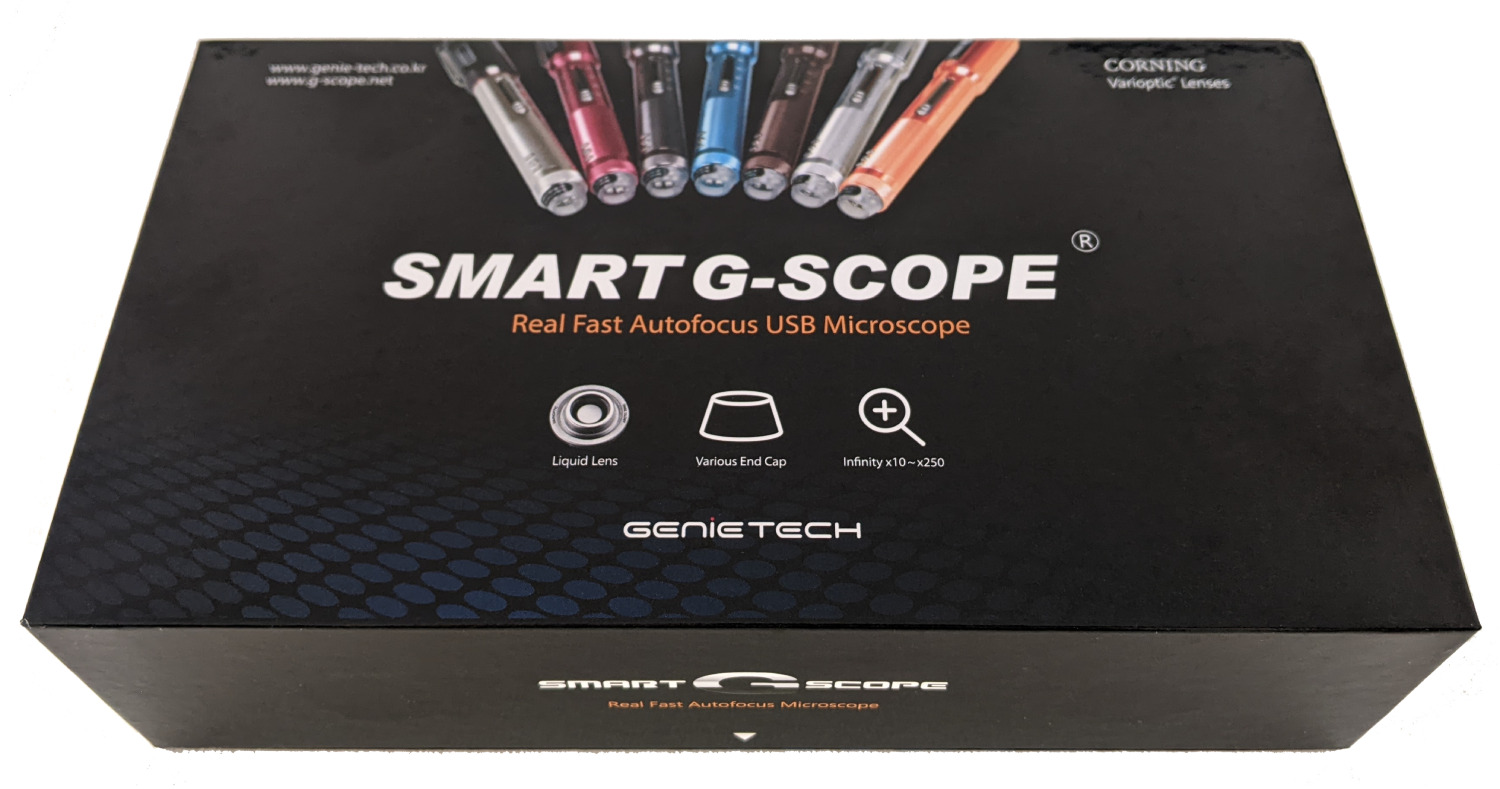
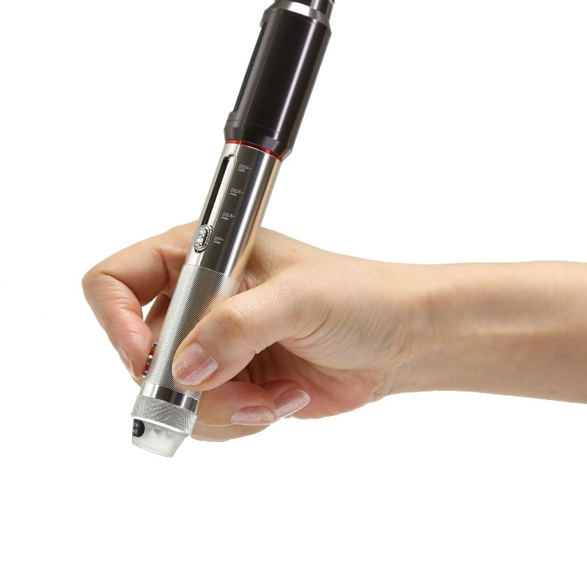
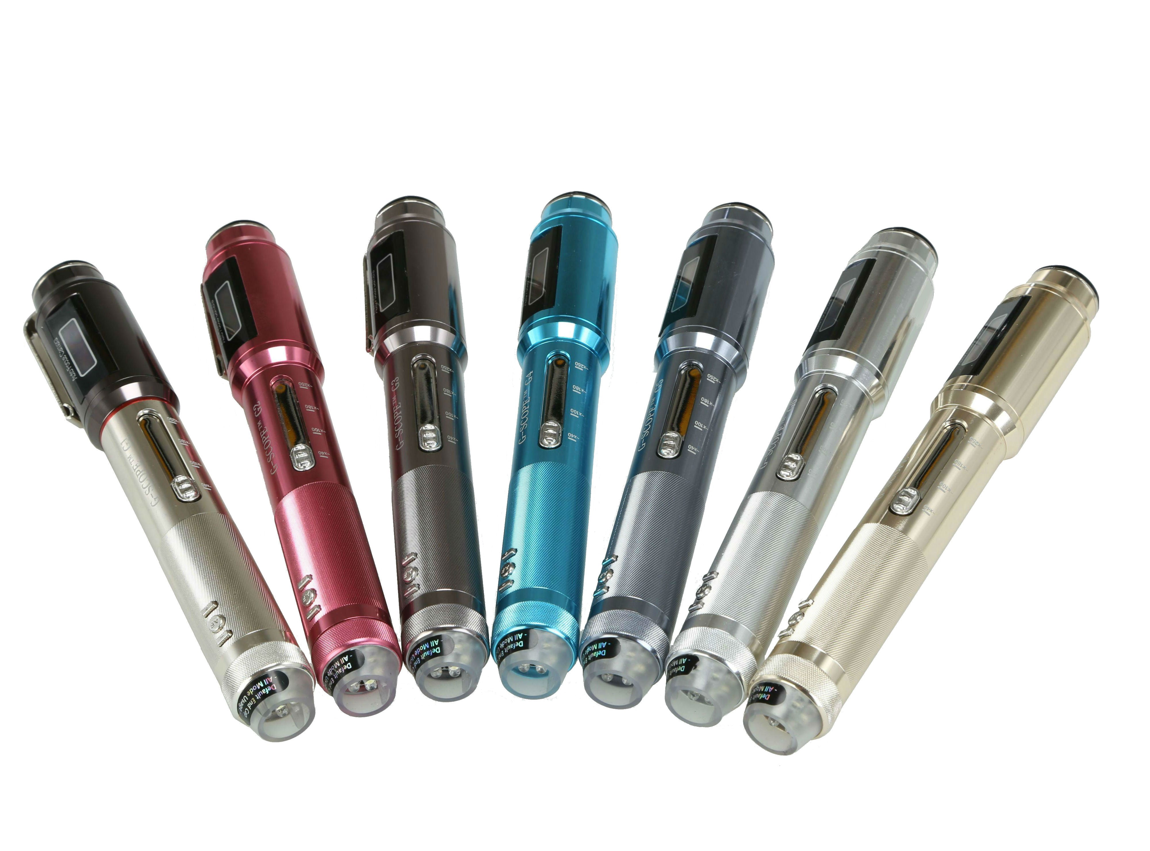
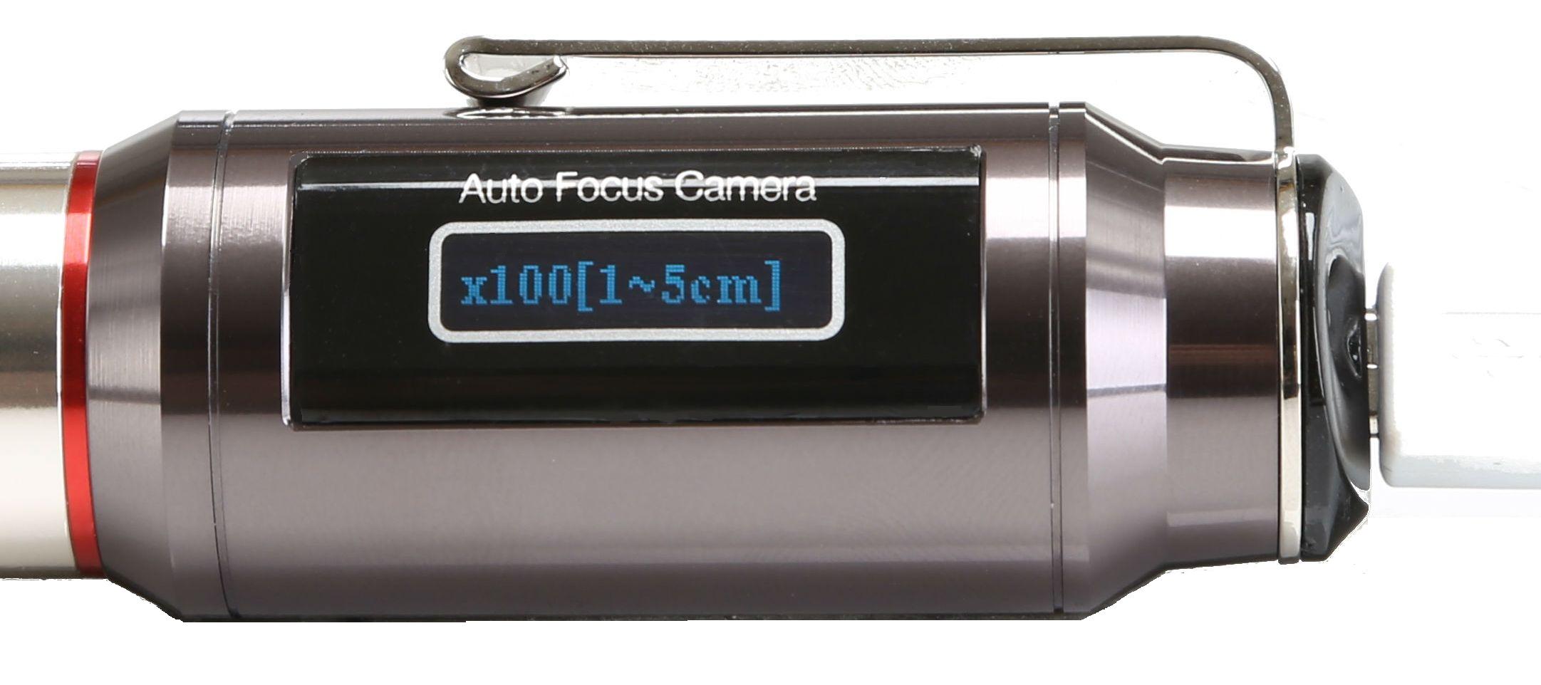
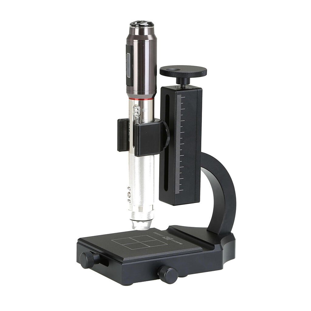
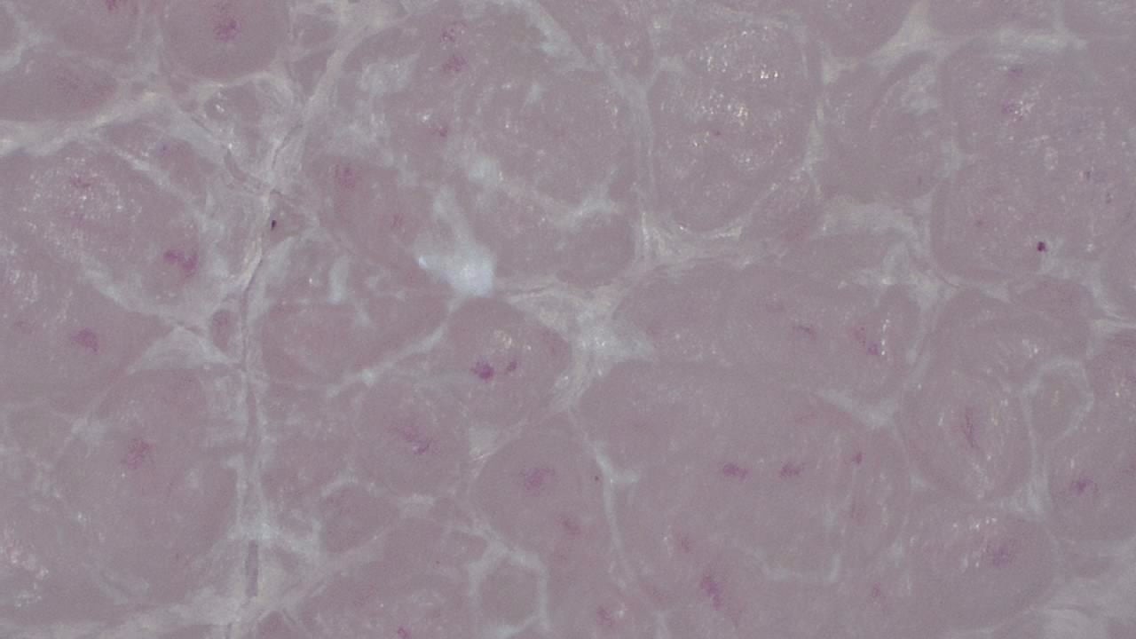
Click any image to enlarge
Practical examples
Onychomycosis follow-up
A podiatrist documents a fungal nail infection over months of topical therapy, showing the patient progressive improvement and supporting adherence.
Plantar wart evaluation
A podiatrist captures images of a plantar lesion for the clinical record. Differentiation, diagnosis, and treatment planning rely on the complete professional assessment.
Limitations & best practices
- Does not replace mycological culture for definitive diagnosis.
- Disinfect the device between patients according to protocol.
- Refer suspicious pigmented lesions to dermatology.